Tools
Anatomy explorer
Turn a springtail round, watch it walk and jump, and tap any part to read what it is. The model is built from the same measurements as the drawing on each species page, and every definition has a source.
The data for the explorer could not be loaded. The drawing below still shows the animal.
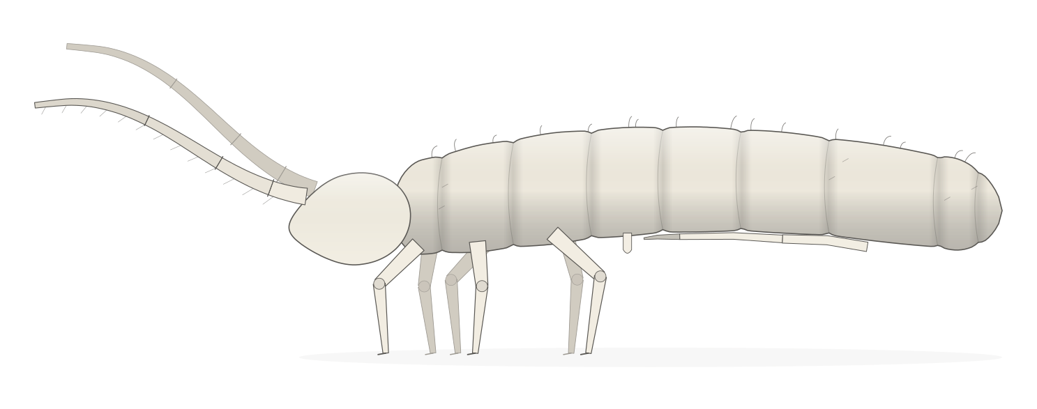
- Folsomia candidaAdult length, 1.2 to 1.8 mm Peer-reviewedOrder Entomobryomorpha
Drag to turn the model. Hold Ctrl and scroll, or click it and scroll, to zoom. Click a part to learn what it is.
Choose a part from the list, or tap it on the model.
Parts
The parts of a springtail
Each part has a glossary entry with its source. In the explorer you can find every one of them on the model.
Head
- Head
- The front part of the body. It carries the antennae, the eyes and the mouthparts. 1 Expert source Open in the glossary (Head)
- Mouthcone
- The snout at the front of the head, formed by the upper and lower lips. It sits low on the front of the head and is flattened rather than truly conical. 1 Expert source Open in the glossary (Mouthcone)
- Eyepatch
- The patch on each side of the head that holds the eyes. It is usually dark, which makes it easy to find with a loupe. 1, 2 Peer-reviewed Open in the glossary (Eyepatch)
- Antenna
- One of the two feelers on the head. Each has four main segments, and some species divide these into many smaller sections. 3 Expert source Open in the glossary (Antenna)
Thorax
- Thorax
- The three body segments behind the head. Each one carries a pair of legs. 3 Expert source Open in the glossary (Thorax)
Abdomen
- Abdomen
- The hind part of the body, made of five segments and a small end piece. It carries the collophore, the retinaculum and the furca. 1, 3 Expert source Open in the glossary (Abdomen)
- Trunk
- The body behind the head. It is the thorax and the abdomen together. 3 Expert source Open in the glossary (Trunk)
- Collophore
- A small tube on the underside of the first abdominal segment with two soft bulbs that can be pushed out. It helps the animal keep its water balance and cling to smooth surfaces. 1, 3 Expert source Open in the glossary (Collophore)
- Anal spines
- Raised spines on the last abdominal segment, near the anus. Some springtails have them and some do not. 1 Expert source Open in the glossary (Anal spines)
Legs
- Leg
- A springtail has three pairs of legs, one pair on each segment of the thorax. From the body outward, a leg is made of two small base pieces, the coxa, the trochanter, the femur and the tibiotarsus, and it ends in the claw. 1 Expert source Open in the glossary (Leg)
- Claw
- The claw at the tip of each leg, called the unguis. Many springtails also have a smaller inner claw or a small pad beside it. 3 Expert source Open in the glossary (Claw)
Furca
- Retinaculum
- A small forked clasp under the third abdominal segment. It holds the folded furca in place until the springtail jumps. 1, 3 Expert source Open in the glossary (Retinaculum)
- Manubrium
- The base piece of the furca. It joins the abdomen at one end and the two arms (dentes) at the other. 1 Expert source Open in the glossary (Manubrium)
- Dens
- One of the two arms of the furca. It runs from the manubrium to the small tip (mucro). The plural is dentes. 1 Expert source Open in the glossary (Dens)
- Mucro
- The small tip at the end of each arm of the furca. Specialists describe its teeth and shape in detail. 1 Expert source Open in the glossary (Mucro)
Surface features
- Tubercle
- A raised bump on the skin, often carrying bristles. Some springtails have prominent tubercles that give them a bumpy, knobbly look. 1 Expert source Open in the glossary (Tubercle)
- Chaeta
- A bristle or hair on the body that sits in a small socket. Their number, length and position help to tell species apart. 1, 2 Expert source Open in the glossary (Chaeta)
- Scales
- Flat, overlapping plates on the body of some springtails. They rub off easily, and they give some species an iridescent shine. 1, 4 Peer-reviewed Open in the glossary (Scales)
What the model is and is not
- The shape is built from the body-plan numbers behind each species drawing. Those numbers are a careful reading of the sources, so the proportions are close but not exact.
- Colour and pattern follow the species notes. Real animals vary from one to the next.
- A size is shown only where a source gives a length. When a length is missing the animals are drawn the same size, and the page says so.
- The walk and the jump are illustrations made for this site. They are drawn slowly and are not measured movements.
- Part names and definitions come from the glossary, which cites its sources. The model adds no claims of its own.
Sources
- 1
Checklist of the Collembola: Glossary of Morphological Terms
Janssens F
Link works · Authoritative database
Cited at s.v. head; s.v. buccal cone; s.v. eye patch; s.v. abdomen; s.v. collophore; s.v. anal spine; s.v. leg; s.v. thorax; s.v. tenaculum; s.v. manubrium; s.v. dens; s.v. mucro; s.v. dorsolateral tubercles; s.v. seta; s.v. scale.
- 2
Katz AD, Giordano R, Soto-Adames F. 2015. ZooKeys 525: 1-75
Verified against a registry · Peer-reviewed paper
Cited at Methods, eye patch setae.
- 3
Checklist of the Collembola of the World
Bellinger PF, Christiansen KA, Janssens F
Link works · Authoritative database
Cited at Introduction, external anatomy; Introduction, etymology.
- 4
Zhang F, Sun DD, Yu DY, Wang BX. 2015. Scientific Reports 5(1)
Verified against a registry · Peer-reviewed paper
Cited at Introduction.